Clinical research software · HUG Geneva
SEEG analysis, simplified.
From raw scans to precise electrode localization —
the complete workflow in one tool, built for clinicians.
Clinical research software · HUG Geneva
From raw scans to precise electrode localization —
the complete workflow in one tool, built for clinicians.
Four steps. One workflow.
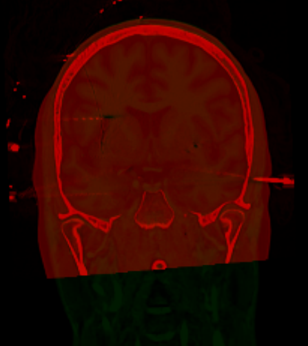
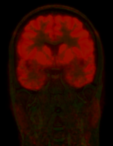
Align MRI, CT, PET and SPECT into a shared space automatically — no scripting, no manual landmarks.
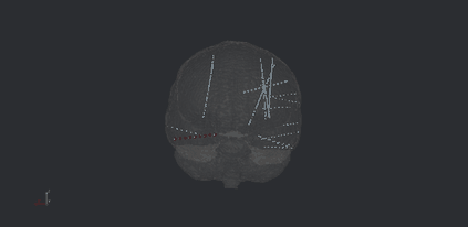
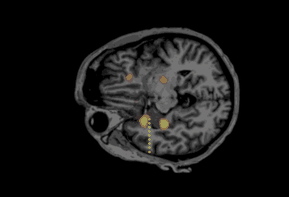
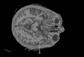
Two clicks per electrode. NeuXelec localizes every contact in axial, coronal and sagittal views.
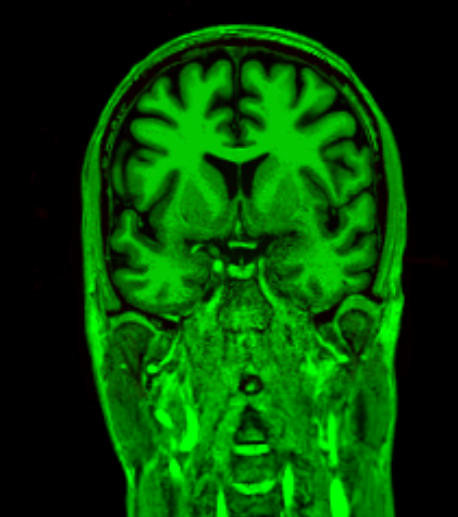
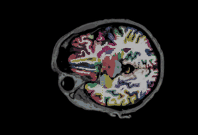
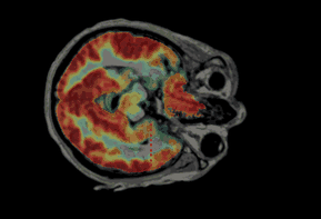
Reslice along the electrode axis. Overlay functional data and map each contact to its brain region.
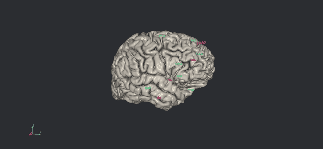
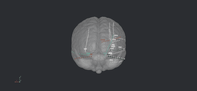
Render all contacts on the cortical surface. Normalize to MNI and export publication-ready data.
Load any combination of T1, CT, T2, PET and SPECT. NeuXelec aligns every modality to the T1 reference automatically — no manual landmark picking, no command line.
Click two contacts on the post-implant CT. NeuXelec reconstructs the full electrode trajectory and assigns precise coordinates to every contact — in minutes, not hours.
Reslice any volume along the electrode shaft for a true contact-aligned view. Overlay CT, MRI, PET and SISCOM simultaneously. A live table maps each contact to its anatomical region.
Visualize all implanted contacts on the patient's cortical surface. Navigate coronal, axial and sagittal slices simultaneously, then normalize to MNI space for group-level analysis.